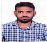
For Emergency Call: 044 – 400 12345

Associate Consultant & Head
Education:
MD, DNB, DM, EBIR
Area(s) of Specialization:
Interventional Neuroradiology, Interventional Vascular Radiology, Development and application of Medical Devices, Computers in Medicine, Imaging Sciences & Physical Principles
Experience:
8 Years of Experience
Consultation days:
Monday to Saturday
Consultation Time:
8 a.m. to 3 p.m.
For Appointment, Call:
044 – 45928653 / EXTN: 8653
PUBLICATIONS AND RESEARCH:
1. SekarSabarish,VinayagamaniS,ThomasB,PoyuranR,KesavadasC.Haemosiderin cap sign in cervical intramedullary schwannoma mimicking ependymoma: how to differentiate? Neuroradiology. 2019 Aug1;61(8):945-8.
2. Sekar Sabarish, Vinayagamani S, Thomas B, Kesavadas C. Arterial spin labeling hyperperfusion in seizures associated with non-ketotic hyperglycaemia: is it merely a post-ictal phenomenon? Neurological Sciences. 2020 Oct12:1-6.
3. Rudrabhatla P, Sabarish S, Ramachandran H, Nair SS. Teaching Neuro Images: A rare adult-onset genetic leukoencephalopathy. Neurology. 2020 Nov18:10-212.
4. Sawant N, Sabarish SS, George T, Shivhare P, Sudhir BJ, Kesavadas C, Abraham M, Jissa VT, Nair P. A Study of the Developing Paediatric Skullbase Anatomy and its Application to Endoscopic Endonasal Approaches in Children. Neurology India. 2020 Sep 1;68(5):1065.
5. Sebastian LJ, Ahuja C, Sekar S, Senthilvelan S, Kulanthaivelu K, Lanka V, Goel V, Mohapatra S, Jain C, Senthilkumaran S, Garg A. COVID-19: Indian Society of Neuroradiology (ISNR) Consensus Statement and Recommendations for Safe Practice of Neuroimaging and Neurointerventions. The Neuroradiology Journal. 2020 Oct;33(5):353-67.
6. Vinayagamani S, Thomas B, Gohil J, Sekar S, Nair P, Kesavadas C. Bipartite craniopharyngeal canal with a lipoma and cephalocele: a previouslyunreported entity. Acta neurochirurgica. 2019 Feb13;161(2):355-9.
7. Vinayagamani S, Sheelakumari R, Sabarish S, Senthilvelan S, Ros R, Thomas B, Kesavadas C. Quantitative Susceptibility Mapping: Technical Considerations and Clinical Applications in Neuroimaging. Journal of Magnetic ResonanceImaging.2020
8. Sabarish S, Kanimozhi P, Nagarajan K. Complex Coronary Cameral Fistulas Evaluated by Multi-Detector CT Angiography: A Report of Three Rare Cases and a Review of the Literature. Cardiovascular Imaging Asia. 2019 Apr1;3(2):47-51.
9. Nagarajan K, Babu AA, Sabarish S, Elango S, Babu KR, Saxena SK. Periorbital Inner Canthal Arteriovenous Malformations: Percutaneous Glue Embolization in Three Cases. Journal of Clinical Interventional Radiology ISVIR. 2020Apr;4(01):55
10. Sunilkumar D, Nagarajan K, Kiran M, Manjubashini D, Sabarish S. Persistent falcine sinus with temporo-occipital schizencephaly: case report with a review ofliterature
in relation to the undeveloped vein of Galen and/or straight sinus. Child's Nervous System. 2019 Jun 1:1-5.
11. Sathiaprabhu A, Sravani N, Nagarajan K, Sabarish S, Patil K. Unusual MR Imaging Features in CT Hyperdense Posterior Fossa Dermoids: Report of Two Cases. Indian Journal of Neurosurgery. 2020Mar;9(01):63-6.
12. Quantitative susceptibility weighted imaging in predicting disease activityin multiple sclerosis- NRAD-D-20-00827R1
13. Teaching Neuroimages: Ohtahara Syndrome Due to Unilateral Perisylvian Polymicrogyria- NEUROLOGY/2020/110965
14. Neuroimaging in CEDNIK syndrome: A Rare Neuro-Ichthyosis, NI-532-19(Inpress)
15. A Novel and distinct pattern of cerebral microbleeds associated with sepsis and respiratory failure presenting as dementia, NI-451-19(Inpress)
16. Maxillo-mandibular vascular malformations: Report of four cases,JCIR
17. Spontaneous intracranial hypotension and lumbosacral spondylolisthesis-case report of a rare association. Clinical Neurology and Neurosurgery. 2021 Jan 20; 202:106511
18. Skeletal complications in congenital insensitivity to pain and anhidrosis: a
problem to reckon with. Neurological Sciences. 2021 Mar 19:1-4.
19. Isolated myelitis and intramedullary spinal cord abscess in melioidosis – a case report, Neurohosptialist (In press)
20. Santhakumar senthilvelan, Sabarish sekar, Chandrasekhran Kesavadas, Bejoy Thomas: Neuromitochondrial disorders- Genomic basis and algorithmic approach to imaging diagnostics-clinical neuroradiology(In press)
21. Joseph JE, Sekar S, Kannath SK, Menon RN, Thomas B. Impaired intrinsic functional connectivity among medial temporal lobe and sub-regions related to memory deficits in intracranial dural arteriovenous fistula. Neuroradiology. 2021 Apr 10:1-9.
HONOURS AND AWARDS:
1. Winner of ABCNR-ISNR Neuroradiology quiz held on 2019, CMC Vellore and won RSNA travel sponsorship award of 1500 dollars and invited interventional neuroradiologyfellowship
2. Winner of ABCNR-ISNR Neuroradiology GRAND JEOPARDY quiz held on New Delhi- 2018 and won RSNA travel sponsorship award of 1500dollars.
3. Second prize in ABCNR Grand quiz, 2017,Mumbai
4. Best Poster award in STAR NEURORADIOLOGY conference held on Pune, August 2017
5. Winner of STAR NEURORADIOLOGY quiz competition held on Pune, August2017.
6. Gold medal in Paediatrics and certificate of merit in Generalmedicine
7. Best poster award, BILIVE – CME on HepatoBiliary Interventions, 2017,Pondicherry
8. First prize in Quiz, BILIVE – CME on HepatoBiliary Interventions, 2017,Pondicherry
CONFERENCES AND CME PRESENTATIONS
1. IRIA 2017, JAIPUR: Benefit of additional ARFI imaging to conventional ultrasound techniques in detection of malignant thyroid nodules- Oralpaper
2. Dense Dermoid Cyst of Posterior Fossa- Unusual imaging features, Best paper award in STAR Neuroradiology conference, Pune, August2017
3. Hydrogen peroxide poisoning- rare cause of Portal venous Gas, Best E-poster award, BILIVE – CME on HepatoBiliary Interventions, 2017,Pondicherry
4. Feasibility of synthetic MRI as clinical tool in various Brain pathologies- ISNR 2018, ISNR, New Delhi
5. Neuroimaging findings of CEDNIK syndrome- ePOSTER - ISNR 2018, NewDelhi
6. Quantitative Susceptibility Weighted Imaging (SWI): A Novel Imaging Biomarker to Predict Disease Activity in Multiple Sclerosis: Oral paper, RSNA Chicago,2019
7. Novel MRI Imaging Techniques in Classification and Evaluation of SpinalVascular Malformations: A Pictoral Review: Education exhibit, RSNA Chicago,2019
CURRENT RESEARCH AND PROJECTS:
1. Resting state functional magnetic resonance imaging in intracranial dural arterio- venousfistula
2. Role of ASL and DTI in characterization of the pituitarymacroadenoma 3. Clinical and angio-graphic predictors of spinal dural arterio-venous fistula